When pregnancy arrives, many fears and fears assail us, perhaps the greatest of which is whether the baby will be developing well. One of the tests that help determine the chances of healthy growth of the fetus is the measurement of the nuchal fold or the verification of the nuchal translucency through an ultrasound.
Percentile tables have been established that correlate the size of the embryo, the weeks of gestation and the thickness of the nuchal translucency, and if on ultrasound there are results that leave the average measurements, it could be a sign that something is not working well. It is known that fetuses with certain chromosomopathies and malformations, such as Down syndrome or cardiac malformations, have a greater nuchal translucency thickness.
Therefore, it is a fundamental test to rule out these problems and, in case of suspicion, to continue doing more specific tests that rule out or confirm the malformations. But what exactly is measured in this test?
Nuchal translucency is the accumulation of fluid in the neck and back of the neck of the fetus, under the skin. Through an abdominal ultrasound this liquid can be easily measured, approximately between the 11th and 14th week of pregnancy. It is usual therefore that the gynecologist perform this test on ultrasound at 12 weeks, together with the test known as "triple screening".
All babies have some fluid in the back of the neck, but fetuses that have an extra chromosome have more fluid in the so-called “nuchal fold”According to the Spanish Society of Gynecology and Obstetrics (SEGO), with high resolution equipment and abdominal probe it is possible to measure translucency or nuchal fold between 10-14 weeks in 95% of fetuses. Its application is equally possible in twin gestations.
 In Babies and more Pregnancy tests: detection of chromosomal abnormalities
 Thanks to the combination of the measurement of the nuchal fold and the triple test, more than 85% of malformations will be detected, the most frequent and known of which are Down, Edwards and Patau syndrome.
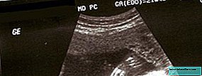
Regarding the technical aspects, you must have an appropriate team and a professional trained and experienced in this measurement task. The cranio-caudal length must be measured with a good sagittal plane of the spine (the fetus must be "caught" on the side, so that a longitudinal section can be seen). The fetus should occupy at least 3/4 of the screen and, if possible, extend the spine and ensure that the amniotic membrane is not attached to the fetal back. Several measurements are made taking the most of them as valid.
When the nuchal fold measurement is performed

The measurement of the nuchal fold is usually performed in the 12th week of pregnancy, on the first abdominal ultrasound. Research has established that nuchal translucency is most effective between weeks 12 and 13.
In this ultrasound, which is usually performed in weeks 11-13, in addition to measuring the nuchal fold, the status of the placenta, the number of fetuses are checked and the gestational age is revealed. This, like any ultrasound in pregnancy, is performed on women to see the evolution of embryonic and fetal structures throughout pregnancy.
 In Babies and more Triple Screening in pregnancy: what to expect from the test
According to gynecological studies, to guarantee the quality of the ultrasound scan, it must be performed by sonographers with experience in measuring the thickness of the TN and equipped with high resolution equipment.
The test works thanks to the conversion of the high frequency sound waves that bounce in the uterus and makes it possible to see an image of its interior and the fetus and it is also possible to see this small space in the neck whose thickness gives us clues about the state of health of the fetus. But, To make the test more effective, its results are combined with another test that is performed at this time of pregnancy, triple screening.
And is that the combination of nuchal translucency and biochemistry offers superior results to those of nuchal translucency as a single method, when both are combined at maternal age, in the prenatal detection of chromosomopathies, both for trisomy 21 and for 18, 13 and monosomy X.
Triple screening and measurement of the nuchal fold

The triple screening test, also called triple test or prenatal screening, is a screening or tracking test (in English, screening) that is done to the pregnant woman to detect possible genetic alterations of the fetus.
It is a non-invasive test (it is made from a sample of maternal blood) that is performed in the first trimester of pregnancy and that consists of an assessment of the risk of chromosomopathy that is obtained by combining three biochemical markers present in the mother's blood :
- PAPP-A (alpha-fetoprotein, protein produced by the fetus)
- Beta-free HCG (human chorionic gonadotropin, the pregnancy hormone, produced by the placenta)
- Free estrogen (estrogen produced by the fetus and placenta)
These biochemical values intersect with the data from the nuchal translucency measurement. of the fetus determined by ultrasound and are weighted according to demographic data such as the mother's age, weight, if she is a smoker, or has diabetes ... Prenatal screening is based on three pillars: mother's age (risk increases with age ), blood tests and nuchal translucency of the fetus.
In the end, a control algorithm is issued that measures the chances that the fetus has an anomaly. According to gynecologists, through this procedure it is possible to detect more than 85% of fetuses at risk of chromosomopathy.
It is estimated that the detection rates can reach 72.7% by applying the combination of Age + nuchal translucency and 86.4% for the combination of Age + TN + Biochemistry, with a false positive rate of 5%.
In any case, it is not a conclusive test, it does not diagnose the malformation but it offers a risk index that the fetus has certain chromosomal alterations such as trisomy 21 (Down Syndrome), trisomy 18 (Edwards Syndrome) and defects of the neural tube.
That is to say, it is a screening test, since it allows the population to be filtered (pregnant in this case), selecting the ones that are most at risk of anomalies, to continue making them diagnostic tests. Here we leave the link to know how to interpret the values of triple screening, although remember to consult all your questions to the specialist.
Normal values of the nuchal fold
The nuchal translucency (TN) is measured between 11 and 14 weeks, because it is at 14 weeks when the thickness of it has reached its maximum, decreasing later. The maximum value of the nuchal fold is usually close to 3 millimeters in normal pregnancies.
The most accepted and internationally used technique is the one proposed by the Fetal Medicine Foundation in London, the organization that sets the international standards, which has drawn up tables with the normal measures of TN in embryos with a maximum embryonic length.
The greater the thickness of the TN, the greater the risk of complications: chromosomal alterations, fetal death and severe malformations. However, we have already pointed out that screening is based on more factors. Therefore, it can happen that the baby has an increased translucency, but the risk index is normal (it can also happen on the contrary).
It should be noted that, according to the Spanish Society of Gynecology and Obstetrics (SEGO), after the review of different authors who publish data on the measurement of the thickness of the nuchal translucency, detection rates and false positives vary widely according to these authors, which may be due to the different quality or experience in ultrasound scanning for this purpose.
Science has shown that, any quantity greater than 3.0 mm is associated with possible genetic malformations.
The risk of anomalies increases with the increase in thickness of the TN: if it is 3-4 mm the risk is 10%, if it is 4-6 mm the risk is 40%, if the risk is greater than 6 mm increases to 80%Ultrasound with increased nuchal translucency

In the event that nuchal translucency gives wide results and there is risk, a detailed ultrasound will be performed by 15 weeks of pregnancy. This ultrasound will look for markers of chromosomopathies and rule out malformations, especially cardiac.
If the specialist considers it necessary, other factors such as the size of the nasal bone can also be evaluated (this is absent in 60-70% of fetuses affected by trisomy 21, about 50% of fetuses with trisomy 18 and in 30 % with trisomy 13).
A genetic study or karyotype of the fetus can also be performed for high risk rates, by amniocentesis or corial biopsy. You have to be aware that the thicker the translucency or nuchal fold, the worse the prognosis.
Undoubtedly, this is one of the situations in which no mother would want to meet and it will surely be a few hard weeks, but fortunately only a very low percentage of pregnancies will confirm a serious problem.
Photos | iStock
More information | Infogen
In Babies and more | What is the measurement of the nuchal fold ?, Tests in pregnancy: detection of chromosomal abnormalities, How to prevent congenital anomalies