It is impossible not to be surprised at the creation of life, almost miraculously two cells that join become a human being in 40 weeks (strictly 38 weeks from fertilization).
I want to share you an animated graphic about the formation of the baby inside the womb in which you can see the different phases of human development from fertilization until birth.

The graph shows the evolution of the baby during 38 weeks from the moment of fertilization, although we see that it covers until week 42 to leave a margin of calculation in case fertilization could have occurred later.
In the first four weeks, the embryonic stage, is in which more changes occur.
First week of pregnancy
When the sperm penetrates the ovum, fertilization and zygote formation (first fertilized cell) occurs. In 72 hours the zygote becomes morula (segmentation of the zygote) and four or five days after fertilization, the morula becomes blastocyte (or blastula).
The blast is composed of two groups of cells, one external and one internal. The internal group will become the embryo, and the outside, the membrane that will protect and nourish it during pregnancy. After the blast is implanted in the endometrium, it is when you start talking about an embryo.
When the blast reaches the uterus, usually six or seven days after fertilization, it begins to produce extensions that will allow it to adhere to the uterine mucosa and "bury" in the endometrium. It is what is known as embryonic implantation.
Embryonic layers
In the graph we can see the three embryonic layers that will give rise to the different tissues and organs of the baby's body.
He ectoderm It is the outermost layer of cells surrounding the embryo: it forms the epidermis, the central and peripheral nervous system, the retina and other structures (hair, nails, tooth enamel).
He endoderm It is the innermost layer, which arises from the first and forms the epithelial coatings of the respiratory ducts and digestive system, including the glands that flow into it. Also bladder, urethra, liver and pancreas.
He mesoderm it originates through the mitosis process of the ectoderm; It is a third layer of cells, located between the ectoderm and the endoderm. It includes smooth muscle layers, connective tissue, vessels that water tissues and organs, blood cells, part of the cardiovascular system, bone marrow and skeleton, striated muscles, and reproductive and excretory organs.
From the second month of pregnancy
From the 5th week of pregnancy (third week of pregnancy) the trilaminar period when some mesodermal cells will give rise to blood vessels that will connect with the embryo circulation through the umbilical vessels, establishing the feto-placental circulation: it is the beginning of the functioning of the cardiovascular system.
In the images of embryos of this gestational age more physical characteristics are already differentiated. On the back you can see a kind of crest that, when closed on itself, forms the neural tube.
On both sides of the neural tube that runs through the back, blocks of tissue called "somites" appear. From them will come the muscles and other body structures. The neural crest will give rise to numerous hundreds and important structures of the embryo: Schwann cells, meninges, melanocytes, adrenal gland marrow or bones.
After the first trimester of pregnancy
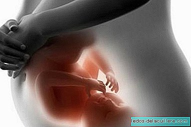
At the end of the first trimester the main structures of the fetus have already been formed. The graph shows the main milestones of baby development such as the formation of the fingers and toes, sexual organs and eyelashes.
From the 24th week of pregnancy you have a 50 percent chance of survival if you were born. In any case, it will continue to form for six months until its development is complete and it is ready to be born (in theory) in week 40.